
This so-called autophagosome is directed to the lysosome where it fuses with the lysosomal membrane and its contents are degraded. In this process, a phagophore membrane is recruited through p62's interaction with LC3 and elongated until it encloses the cargo-p62 complex in a double-membrane vesicle. Due to its early discovery, p62 can be considered the archetypical autophagy receptor that is also involved in the targeted removal of other cargo types such as bacteria, viruses, and organelles ]. The molecular link to autophagy was established by p62's involvement in the disposal of poly-ubiquitinated aggregates via the lysosome ]. Further interaction studies revealed that p62 bridges the interaction of atypical protein kinase C (PKC) with RIP1 to activate the NF-κB pathway ]. Approximately 25 years ago, p62 was identified as a novel interaction partner of the SH2 domain of tyrosine–protein kinase Lck and subsequently cloned ].
#Framework protein scaffold series#
P62/SQSTM1 (from here on p62) is a multidomain, multifunctional protein involved in autophagy and a series of signaling processes ]. fluorescence recovery after photobleaching.Abbreviations used: ABD albumin-binding domain of protein G APPI Alzheimer's amyloid beta-protein precursor inhibitor BBP bilin-binding protein BPTI bovine (or basic) pancreatic trypsin inhibitor BSA bovine serum albumin CBD cellulose-binding domain of cellobiohydrolase I CD circular dichroism Cdk2 human cyclin-dependent kinase 2 CDR complementarity-determining region CTLA-4 human cytotoxic T-lymphocyte associated protein-4 FN3 fibronectin type III domain GSH glutathione GST glutathione S-transferase hIL-6 human interleukin-6 HSA human serum albumin IC(50) half-maximal inhibitory concentration Ig immunoglobulin IMAC immobilized metal affinity chromatography K(D) equilibrium constant of dissociation K(i) equilibrium dissociation constant of enzyme inhibitor LACI-D1 human lipoprotein-associated coagulation inhibitor pIII gene III minor coat protein from filamentous bacteriophage f1 PCR polymerase-chain reaction PDB Protein Data Bank PSTI human pancreatic secretory trypsin inhibitor RBP retinol-binding protein SPR surface plasmon resonance TrxA E. An overview will be provided about the current approaches, and some emerging trends will be identified.
#Framework protein scaffold how to#
This review will therefore concentrate on the critical description of the structural properties of experimentally tested protein scaffolds and of the novel functions that have been achieved on their basis, rather than on the methodology of how to best select a particular mutant with a certain activity. However, it appears that not all kinds of polypeptide fold which may appear attractive for the engineering of loop regions at a first glance will indeed permit the construction of independent ligand-binding sites with high affinities and specificities. Recently, the scaffold concept has even been adopted for the construction of enzymes. Hence, among others, single domains of antibodies or of the immunoglobulin superfamily, protease inhibitors, helix-bundle proteins, disulphide-knotted peptides and lipocalins were investigated.

Properties like small size of the receptor protein, stability and ease of production were the focus of this work. After the application of antibody engineering methods along with library techniques had resulted in first successes in the selection of functional antibody fragments, several laboratories began to exploit other types of protein architectures for the construction of practically useful binding proteins.


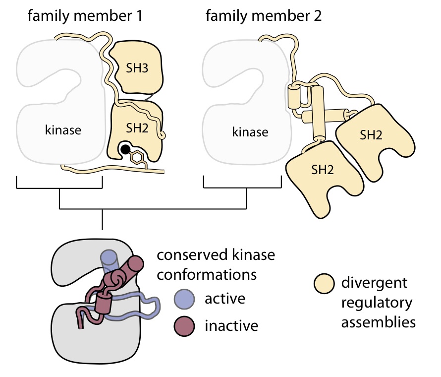
This development started with the notion that immunoglobulins owe their function to the composition of a conserved framework region and a spatially well-defined antigen-binding site made of peptide segments that are hypervariable both in sequence and in conformation. The use of so-called protein scaffolds has recently attracted considerable attention in biochemistry in the context of generating novel types of ligand receptors for various applications in research and medicine.


 0 kommentar(er)
